FDA approves first cardiac use of da Vinci Surgical System after successful trial led by ECU surgeons
GREENVILLE, N.C. (Nov. 13, 2002) — Cardiac surgeons at the Brody School of Medicine at East Carolina University have led a successful national multi-center clinical trial studying the use of the da Vinci Surgical System for mitral valve repairs. This breakthrough robotic surgical procedure was approved for its first cardiac use by the U.S. Food and Drug Administration Nov. 12.
Dr. W. Randolph Chitwood Jr., chairman of the Department of Surgery at East Carolina University, served as principal investigator of the 10-hospital study. He has performed 51 mitral valve surgeries using the da Vinci system – more than any other cardiac surgeon in the world.
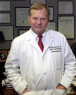
Dr. W. Randolph Chitwood. Photo by Cliff Hollis.
After receiving word of the authorization Tuesday, Chitwood performed the first post-approval mitral valve repair using the robotic device on a 74-year-old woman from Idaho Falls, Idaho. She is recovering well, Chitwood said.
The multi-center FDA trial involved 112 patients, with ECU enrolling 22 patients. All ECU patients in the trial had their surgeries performed at Pitt County Memorial Hospital, the affiliated teaching hospital of the medical school. Before participating in the trial, surgeons at the nine other medical centers received training using da Vinci for this procedure through the Minimally Invasive Surgery/Computer Enhanced Training Center at ECU, the only site in the world offering certified mitral valve training on the robotic device.
The clinical trial studied the safety and efficacy of the minimally invasive, robotic-assisted technique versus traditional sternotomy, or open-chest procedure, in repairing defective mitral valves. Cardiothoracic surgeons use mitral valve repair to treat a narrowing or leakage of the mitral valve. The mitral valve has two leaflets, or flaps, and allows blood to flow from the heart”s left atrium into the left ventricle. When the valve leaks, blood backs up into the lungs and causes the ventricle to pump more blood. Symptoms include irregular heart rhythms, shortness of breath and fatigue.
Traditional open-heart surgery requires surgeons to make an 8- to 10-inch incision by sawing through the sternum and opening the rib cage to gain access to the heart.
With the da Vinci Surgical System, Chitwood and his colleagues make a 4-centimeter incision between the ribs in the right chest and two other 1-centimeter incisions before inserting three robotic arms into the chest. One arm holds a tiny camera that projects 3-D images onto a monitor in front of the surgeon. The lens system magnifies millimeter-sized arteries and veins 10 times, to the size of drinking straws. The other two arms hold the pencil-sized instruments, which have tiny computerized mechanical “wrists” designed to transmit the dexterity of the surgeon”s forearm and wrist into the chest at the operative site.
Seated at a computer console located apart from the operating table, the surgeon views a magnified, 3-D image while manipulating the surgical instruments using two joystick-like devices.
“Clearly there is a shorter learning curve with the da Vinci Surgical System than with standard laparoscopic surgery,” Chitwood said. “The operative times fall very rapidly within the first 20 cases. These operations still are somewhat longer than conventional breast-bone incision operations, but proven data suggests improvement in the patients’ recovery with less complications in the selected patients upon whom we have operated thus far for mitral valve disease.
“A continuum of improvements could make robotic telemanipulation supplant conventional mitral valve surgery; however, it will require wide acceptance of surgeons who will look for facilitating adjunctive technologies to apply with the robot method.”