ECU students and faculty work to explain blood clot formation and disease
East Carolina University’s Dr. Nathan Hudson is leading research to better understand how blood clots form, what impacts degradation of clots and new ways to attack them to help improve the diagnosis and treatment of disease. He also is developing new materials that can aid medical professionals in surgery and wound healing procedures.
Hudson is an associate professor in the Thomas Harriot College of Arts and Sciences Department of Physics and studies the exciting field of molecular biophysics and biomedical physics. Science and medicine work hand-in-hand in these fields, and specialists examine biological samples and living systems through a quantitative, physical science-inspired lens to better understand biomedical processes.
Many of Hudson’s research projects have received federal grant funding to help solve fundamental questions about the formation and degradation of blood clots, which may lead to the development of innovative compounds capable of treating disease.
His newest project is funded by a $450,000 National Heart, Lung and Blood Institute (NHLBI) grant from the National Institutes of Health. Collaborators include ECU students; Dr. Martin Guthold, a professor of physics at Wake Forest University; and Dr. Brittany Bannish Laverty, a professor and mathematician at the University of Central Oklahoma.
“For decades people have studied the structures of fully formed blood clots, and we now know there are strong connections between blood clot structures and a person’s health,” Hudson said. “However, blood clots form so quickly that we have large gaps in our understanding of what regulates the final clot structure and how this is altered in diseases.”
Students assist in research

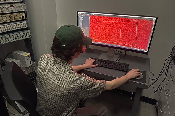
Physics major Dylan Miller analyzes data of a forming blood clot. (Contributed photos)
To help answer these questions, integrated biomedical physics doctoral student Aravind Elangovan and undergraduate physics major Dylan Miller are working with Hudson using a new microscopy technique, known as light sheet microscopy, that can capture movies of blood clots while they are forming.
This method illuminates a thin portion of a sample, as opposed to the entire sample. Therefore, the sample is less damaged by the light and can be used for a longer time, while the researcher collects data much quicker.
“Research is exciting because you are in the frontier of your field. The distant future of this research is tangible,” Elangovan said. “Many diseases affect how a blood clot behaves, or if it even forms in the first place. So, learning about how it forms will give insight into its behavior. If we know how something behaves, then we can fix it.”
Elangovan said he is gaining beneficial lab experience and some computer programming skills, which will assist him in his future medical physicist career.
“I like the versatility of it,” he said.
Hudson said that understanding how blood clots form has implications for human health.

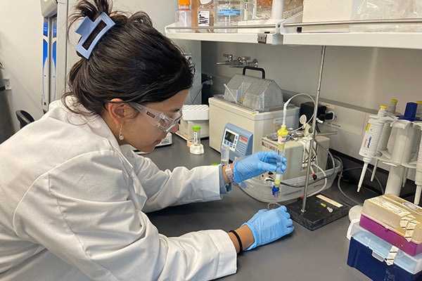
Biology major Maria Gonzalez Mundarain uses an affinity column to purify fibrinogen from human plasma.
“Pathologies such as strokes and heart attacks often occur because blood clots aren’t digested properly,” he said. “We intend to connect the process of blood clot formation with the process of digestion.”
Another important aspect of Hudson’s research is student training.
“Students develop skill sets in physics, biochemistry, mathematics, computer science, engineering and medicine,” he said. “Such an interdisciplinary skill set is invaluable in the competitive job market and sets students up for success in the future.”
Undergraduate students Victoria Gonzalez Mundarain, a biology and biochemistry major, and Maria Gonzalez Mundarain, a biology major, are leading investigators on another of Hudson’s NIH-funded NHLBI grants. They are working on developing molecular structures of blood clotting proteins in solution, and they have demonstrated the blood clotting protein fibrinogen is much more flexible than was previously believed. The long-term goals of this project are to develop molecular structures that reveal the early stages of the blood clotting process and discover new targets for drug design.
